Some organelles are easily viewed with a light microscope.
Biology Microscopy - Organelles
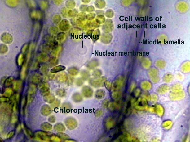
Anacharis (Elodea) leaf cells x1000.
This plant is sold in pet stores for use in home fish aquaria. Green chloroplasts appear mottled because of their internal grana. In this specimen you can see a nucleus and nucleolus, something not often seen in such cells.
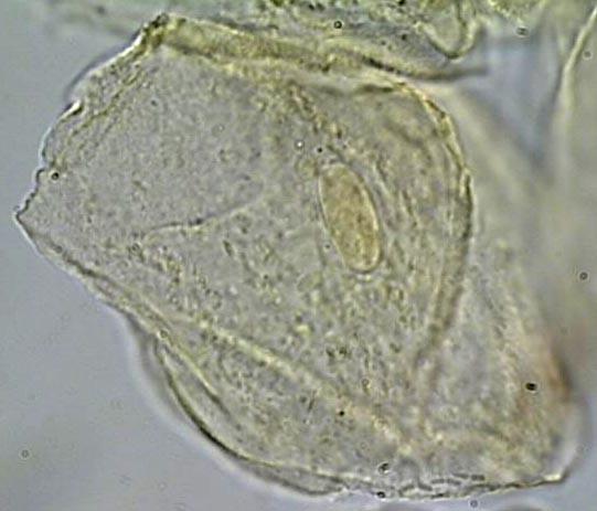
Human cheek cell x1000.
The cells have been stained with to make them easier to see. The oval nucleus of this very flat cell is quite noticeable.
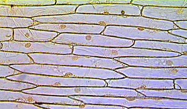
Onion epidermis x100.
Each cell has a nucleus. The specimen has been stained with gram iodine to give the nuclei more contrast.
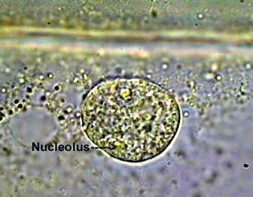
Onion epidermis nucleus x1000.
One of the three nucleoli visible in the nucleus is labeled.
Potato cells with unstained leucoplasts x100.
Leucoplasts are the egg-shaped structures.
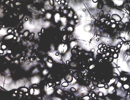
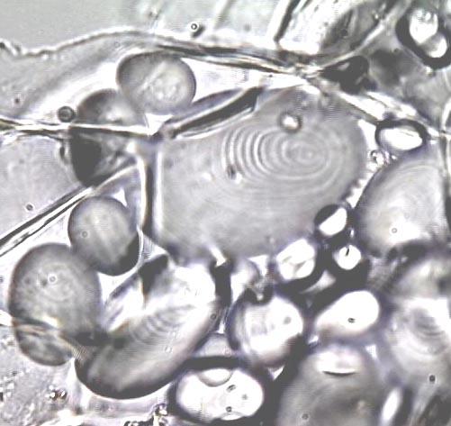
Leucoplasts x1000.
These are unstained leucoplasts in potato cells.
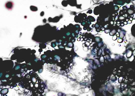
Potato cells with stained leucoplasts x100.
The leucoplasts have been stained with gram iodine. The resulting purple, blue, or black color is chemical proof that leucoplasts contain starch.